Are you experiencing sudden and intense pain in your joints? Wondering if it might be gout? Well, fret not, because in this article we will explore the various diagnostic methods and tests used to identify and confirm gout. From analyzing your symptoms to conducting blood tests, this will be your go-to guide to understand how gout is diagnosed. So, let's dive into the world of gout diagnosis and find out what steps you need to take to get a clearer picture of your condition.
Medical History
When it comes to diagnosing gout, one of the first steps is to gather a comprehensive medical history from the individual in question. This includes information about past and current symptoms, family history, and any relevant medical conditions or medications the individual may be taking. By obtaining this information, healthcare professionals can gain important insights that will help guide the diagnostic process.
Symptoms
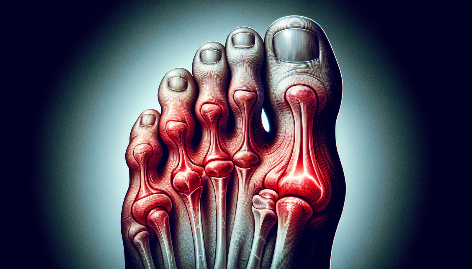
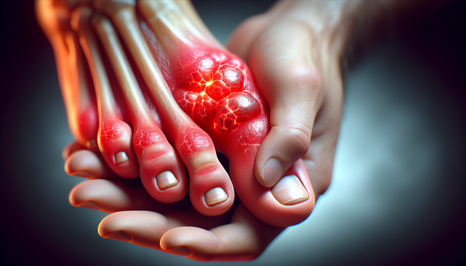
In order to make an accurate diagnosis of gout, it is crucial to assess and understand the various symptoms that typically accompany this condition. Gout is known for causing sudden and severe joint pain, most commonly in the big toe, although it can also affect other joints such as the ankles, knees, and wrists. The pain experienced during a gout attack is often described as throbbing or excruciating, and may be accompanied by redness, swelling, and warmth in the affected area.
Family History
Another key aspect of the medical history that plays a significant role in the diagnosis of gout is the patient's family history. Gout has a genetic component, so individuals with a family history of gout are at an increased risk of developing the condition themselves. By understanding if there is a family history of gout, healthcare professionals can further solidify their suspicions and potentially expedite the diagnostic process.
Physical Examination
After gathering the necessary medical history, a physical examination is typically conducted to evaluate the individual's symptoms and assess any physical signs of gout. This examination focuses on two main areas: joint examination and skin examination.
Joint Examination
During a joint examination, the healthcare professional will carefully assess the affected joints for signs of inflammation, such as redness, swelling, and tenderness. In gout, these symptoms are typically localized to one or a few joints and may be accompanied by limited range of motion and increased pain upon movement. By conducting a thorough joint examination, healthcare professionals can gather additional evidence to support their suspicion of gout.
Skin Examination
In some cases, gout can manifest with visible skin abnormalities. Therefore, a skin examination is an important part of the diagnostic process. In particular, healthcare professionals look for the presence of tophi, which are small, firm nodules that form beneath the skin due to the accumulation of uric acid crystals. Tophi usually develop in areas such as the elbows, fingers, and ears. Identifying the presence of tophi through a skin examination can be highly indicative of gout and contribute to an accurate diagnosis.
Lab Tests
In addition to the medical history and physical examination, laboratory tests are commonly utilized to support the diagnosis of gout. The two main types of lab tests used for this purpose are blood tests and kidney function tests.
Blood Tests
A blood test can provide valuable information about the levels of uric acid in the body. Uric acid is a waste product that is formed when the body breaks down purines, which are found in certain foods and drinks. In individuals with gout, the level of uric acid in the blood is often higher than normal. However, it is important to note that high levels of uric acid alone do not necessarily confirm a diagnosis of gout, as some individuals may have elevated levels without experiencing any symptoms.
Uric Acid Levels
Measuring uric acid levels in the blood is an important diagnostic tool for gout. Generally, a uric acid level of 6.8 milligrams per deciliter (mg/dL) or higher is indicative of hyperuricemia, which is a condition where excess uric acid accumulates in the bloodstream. While hyperuricemia is not always synonymous with gout, it is a common feature of the condition and can help support a diagnosis.
Kidney Function Tests
Since gout is closely linked to kidney function, kidney function tests may also be ordered during the diagnostic process. These tests assess the overall health and function of the kidneys and can detect any abnormalities or impairments in their ability to filter and excrete uric acid. Abnormal kidney function can contribute to the development of gout and may influence the management and treatment plan for individuals diagnosed with the condition.
Imaging Tests
Imaging tests, such as X-rays, ultrasound, and dual-energy CT scan, are often used in the diagnostic process to provide a visual representation of the affected joints and surrounding tissues.
X-rays
X-rays are commonly used to evaluate joint damage and detect the presence of joint erosions in individuals suspected of having gout. In gout, X-ray findings may reveal characteristic joint erosions, which are areas of bone loss or destruction caused by the deposition of uric acid crystals. These erosions are typically seen in advanced or chronic cases of gout and can aid in confirming a diagnosis.
Ultrasound
Ultrasound imaging can also be a valuable tool in diagnosing gout. It allows for a closer examination of soft tissues and can help identify the presence of tophi, which may not be easily visible during a physical examination. Ultrasound findings showing the presence of tophi in or around the affected joints can contribute to the diagnosis of gout.
Dual-energy CT Scan
A dual-energy CT scan is a specialized imaging technique that can detect the presence of uric acid crystals in and around the joints. This non-invasive test can help visualize and map the distribution of uric acid crystals, providing further evidence to support a diagnosis of gout. Dual-energy CT scans are particularly useful in cases where the diagnosis may be in question, or when traditional imaging methods, such as X-rays or ultrasound, do not provide conclusive results.

Joint Fluid Analysis
One of the most definitive tests used to diagnose gout is joint fluid analysis. This procedure involves the extraction of synovial fluid from the affected joint and subsequent analysis of the fluid sample.
Synovial Fluid Extraction
Joint fluid, also known as synovial fluid, is a lubricating fluid that fills the joint cavity. In individuals with gout, this fluid may contain uric acid crystals, which are the hallmark of the condition. To obtain a sample of the synovial fluid, a healthcare professional will first numb the area around the joint and then use a needle to withdraw a small amount of fluid for analysis.
Microscopic Examination
Once the synovial fluid sample has been obtained, it is sent to a laboratory for microscopic examination. Under a microscope, healthcare professionals can directly visualize the presence of uric acid crystals in the synovial fluid. The identification of these crystals is highly indicative of gout and can confirm the diagnosis.
Chemical Analysis
In addition to microscopic examination, a chemical analysis of the synovial fluid may also be performed. This analysis provides additional insights into the composition of the fluid, including the levels of uric acid present. Abnormally high levels of uric acid in the synovial fluid further support the diagnosis of gout.
Differential Diagnosis
In the process of diagnosing gout, it is essential to rule out other conditions that may present with similar symptoms. This is known as a differential diagnosis.
Ruling out Other Conditions
Gout can sometimes be confused with other forms of arthritis, such as rheumatoid arthritis or pseudogout, which is caused by the deposition of calcium pyrophosphate crystals. By carefully considering the individual's symptoms, medical history, and results of lab and imaging tests, healthcare professionals can eliminate other potential causes and focus on making an accurate diagnosis of gout.
Distinguishing Gout from Similar Conditions
Differentiating gout from other conditions can be challenging due to similar symptoms and overlapping features. However, certain factors, such as the pattern and duration of joint inflammation, the presence of tophi, and the identification of uric acid crystals, can help distinguish gout from similar conditions. This diagnostic process requires meticulous analysis and expertise from healthcare professionals to ensure an accurate diagnosis and appropriate treatment plan.

Classification Criteria
To aid in the diagnosis of gout, medical organizations, such as the American College of Rheumatology (ACR), have established classification criteria that help standardize the diagnostic process.
American College of Rheumatology (ACR) Criteria
The ACR criteria for the classification of gout take into account various clinical and laboratory findings. These criteria include the presence of characteristic joint inflammation, a history of at least one gout attack, the presence of tophi or uric acid crystal deposits, and the detection of uric acid in the synovial fluid. Meeting these criteria adds further confidence to the diagnosis of gout.
Medical Imaging
Medical imaging plays an integral role in the diagnosis of gout, as it provides visual evidence of joint damage and the presence of uric acid crystals.
X-ray Findings
X-rays can reveal joint erosions, typically seen in chronic cases of gout. These erosions occur as a result of the uric acid crystals gradually damaging the bone. X-ray findings showing joint erosions, along with other clinical indicators, can support the diagnosis of gout.
Ultrasound Findings
Ultrasound imaging can demonstrate the presence of tophi, which are collections of uric acid crystals. Tophi may appear as hyperechoic, or bright, areas within the soft tissues surrounding the joints. Detecting tophi on an ultrasound can provide strong evidence in favor of a gout diagnosis.
Dual-energy CT Scan Findings
Dual-energy CT scans are exceptionally useful in diagnosing gout through the identification of uric acid crystal deposits. The scan can visualize the distribution and extent of these crystals, helping healthcare professionals confirm the presence of gout and differentiate it from other conditions with similar manifestations.

Additional Tests
In certain cases, additional tests may be needed to support the diagnosis of chronic gout or to provide further insights into the condition.
Kidney Function Tests
As previously mentioned, gout is closely related to kidney function. Monitoring and evaluating kidney function through additional tests, such as measuring creatinine levels and estimating glomerular filtration rate (eGFR), can help assess the overall health of the kidneys and identify any underlying issues that may contribute to the development or management of gout.
Kidney Stone Analysis
In some instances, individuals with gout may also develop kidney stones. Analyzing the composition of these stones can offer valuable information about the underlying causes and characteristics of the condition. This additional test can aid healthcare professionals in creating a more comprehensive treatment plan for those with kidney stones and gout.
Genetic Testing
While genetic testing is not commonly utilized in the diagnosis of gout, it may occasionally be recommended, particularly in cases where there is a strong family history of the condition. Genetic testing can help identify specific gene mutations or variations that may increase an individual's susceptibility to developing gout. This information can be valuable in understanding the underlying genetic factors involved and may guide treatment decisions or preventive measures.
Diagnosing Chronic Gout
Chronic gout refers to cases where the disease has become recurrent or has progressed to a more advanced stage. There are specific indicators that healthcare professionals look for when diagnosing chronic gout.
Identification of Tophi
The presence of tophi, which are collections of uric acid crystals beneath the skin, is a key characteristic of chronic gout. The identification of tophi during a physical examination or through imaging tests can confirm chronic gout and guide treatment decisions.
Joint Erosions on Imaging
When assessing chronic gout, imaging tests such as X-rays or CT scans may reveal joint erosions, which are areas of bone loss due to the accumulation of uric acid crystals. The presence of these erosions, along with other diagnostic criteria, can confirm the diagnosis of chronic gout and indicate the need for long-term management strategies.
In conclusion, diagnosing gout requires a comprehensive approach that encompasses a thorough medical history, physical examination, lab tests, imaging tests, joint fluid analysis, and consideration of differential diagnoses. By gathering and analyzing all available information, healthcare professionals can accurately diagnose gout and tailor an appropriate treatment plan aimed at managing symptoms and preventing future gout attacks.


